Real-time insight into fresh tissue
Instant cellular and architectural information that supports precise intraoperative decisions and streamlines the diagnostic workflow.
Clinical benefits
Instant cellular and architectural information that supports precise intraoperative decisions and streamlines the diagnostic workflow.
What we deliver
The histological features today’s pathology teams rely on, visualized in minutes.
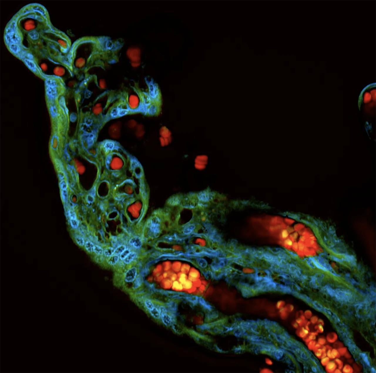
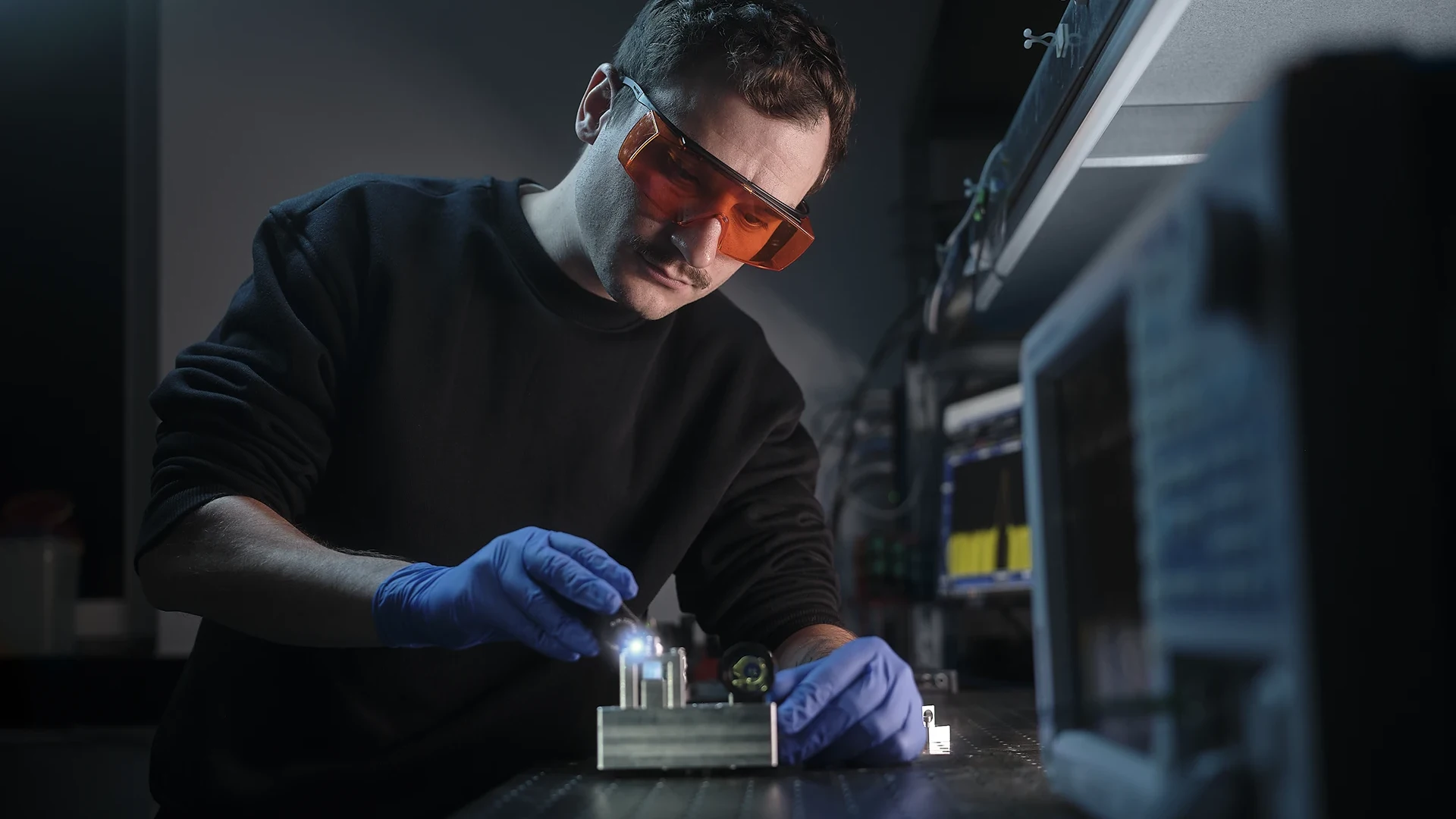
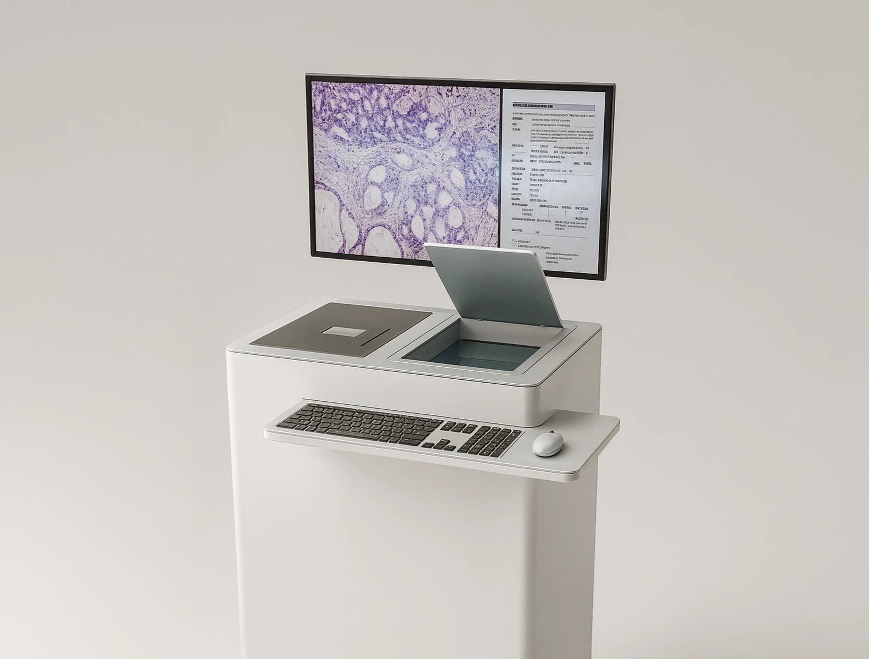
Refined Diagnostics generates virtual histology from fresh, unstained tissue, visualizing cellular morphology and tissue architecture with contrast comparable to standard stains. It also captures structural and metabolic features, including endogenous cofactors like NADH and FAD, in 3D datasets.
Unlike conventional histology, these images are obtained without staining or sectioning. Biopsies can be placed directly into the system for immediate imaging while remaining intact for downstream analyses.
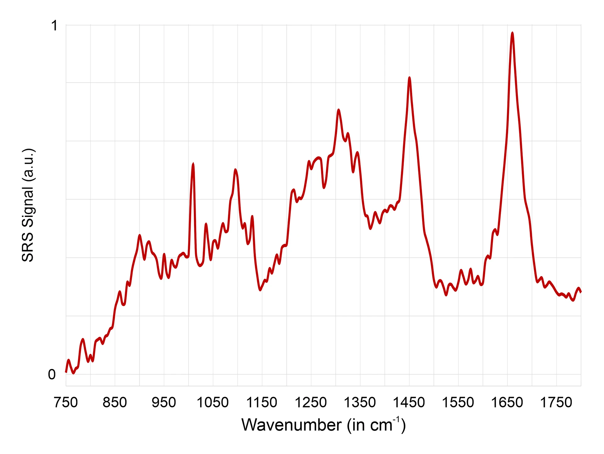
Beyond morphology, the Raman-based molecular fingerprint provides spatially resolved biochemical information that complements established techniques such as IHC, PCR, and DNA analysis, opening new possibilities for tumor classification, biomarker discovery, and AI-driven pathology.
Why it matters
A unified platform for adequacy assessment, surgical guidance, and rapid molecular insight.
For patients with suspicious lung nodules, diagnosis often involves days of uncertainty and repeat biopsies when samples are insufficient. Refined Diagnostics delivers on-site adequacy assessment within minutes, allowing immediate quality confirmation and fast resampling. This reduces anxiety, shortens the diagnostic path, and improves clinical efficiency.
During resections, surgeons often depend on visual judgment and delayed pathology, leading to repeat surgeries when residual tumor is found later. Refined Diagnostics provides real-time visualization of tissue architecture in the OR, supporting precise margin detection and preservation of healthy tissue. This increases confidence and lowers the need for follow-up interventions.
Molecular and immunohistochemical data typically require days to obtain. Refined Diagnostics extracts molecular fingerprints directly from fresh tissue, adding actionable context to intraoperative morphology. This can accelerate diagnostic workflows and enable earlier insights into tumor biology and potential biomarkers.
Workflow
Seamless integration of preparation-free tissue imaging into surgical and pathology workflows for faster insight.
1

Position the fresh, unstained tissue on the imaging stage with no further processing.
2

Aquila acquires Raman and additional signals within minutes, generating depth-resolved data.
3

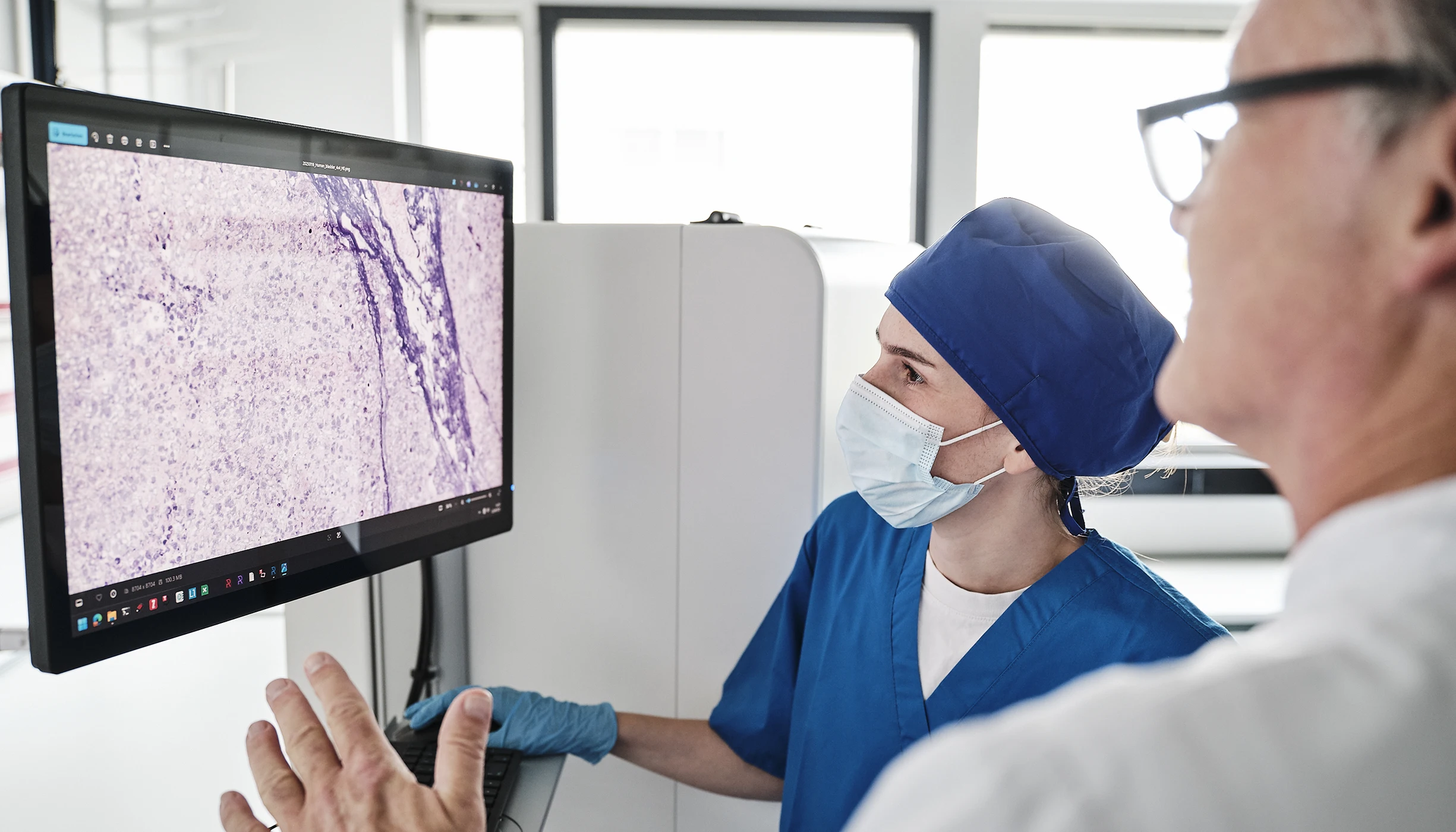
Morpho-molecular details support decisions during surgery and guide diagnostic review.
Refined Diagnostics integrates seamlessly into existing workflows. Fresh, unstained tissue is placed directly into the system with no preparation. Within minutes, high-resolution cellular and structural data are available for review, sharing, and documentation. The three steps—place, image, interpret—provide diagnostic-level insight at the point of care and enable faster, coordinated decisions.
Our solution
Learn more about system specifications, imaging performance, and integration options.
Contact