Label-free imaging with highest contrast
Label-free, multicolor imaging by detecting intrinsic molecular vibrations, revealing lipid metabolism, protein synthesis, and drug dynamics in real time without photobleaching.
SRS imaging solutions
Label-free, multicolor imaging by detecting intrinsic molecular vibrations, revealing lipid metabolism, protein synthesis, and drug dynamics in real time without photobleaching.
Every molecule vibrates in a unique way, creating an optical fingerprint that reveals its chemical identity. Raman microscopy detects these vibrations to visualize the molecular composition of cells and tissues directly, without stains or labels.
Coherent Raman imaging, including SRS and CARS, enhances the signal by several orders of magnitude and enables imaging speeds fast enough to capture biological dynamics in real time.
Benefits
Experience imaging that reveals true molecular detail – fast, clean, and unrestricted by traditional markers.
Dye-free imaging of living cells and fresh tissue, preserving the native biological state.
Captures chemically specific information, enabling data-rich analysis beyond morphology.
Subcellular resolution revealing fine structural and molecular details.
Enables real-time observation of living cells and dynamic biological processes without causing damage.
Molecular depth
Reveal subtle chemical patterns, material signatures, and physiological processes instantly – delivering deeper understanding without adding steps or dyes.
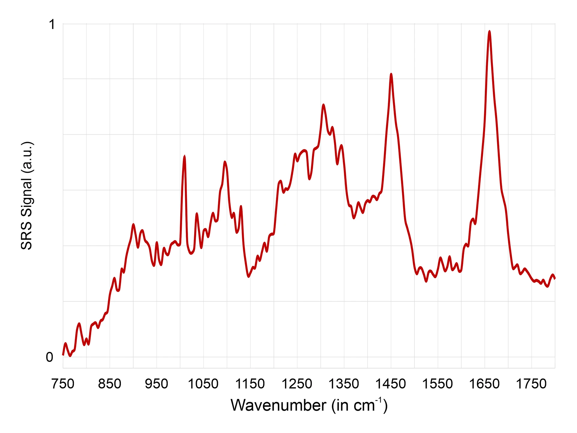
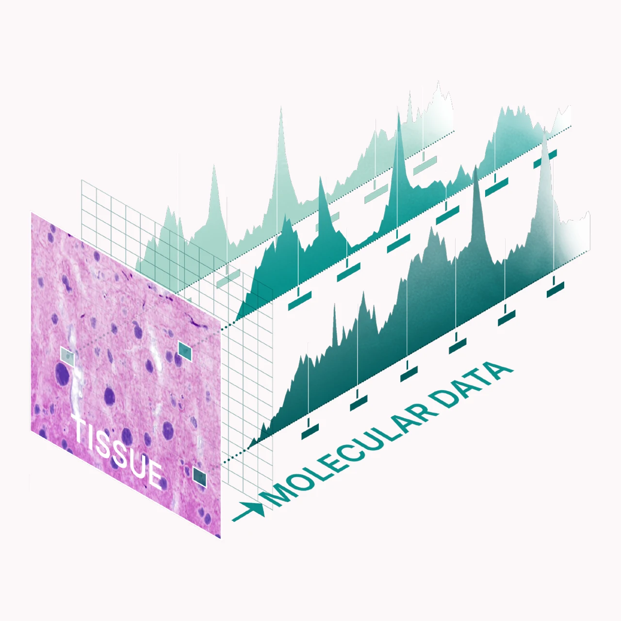
Each SRS pixel contains a full molecular spectrum that reveals local chemical composition. These vibrational signatures from lipids, proteins, and nucleic acids provide chemical contrast beyond structure alone. Capturing spectra across the sample enables label-free biochemical imaging. With high tuning speed and a compact design, Refined Laser Systems records these molecular fingerprints rapidly and reliably, making advanced SRS imaging practical for real biological research.
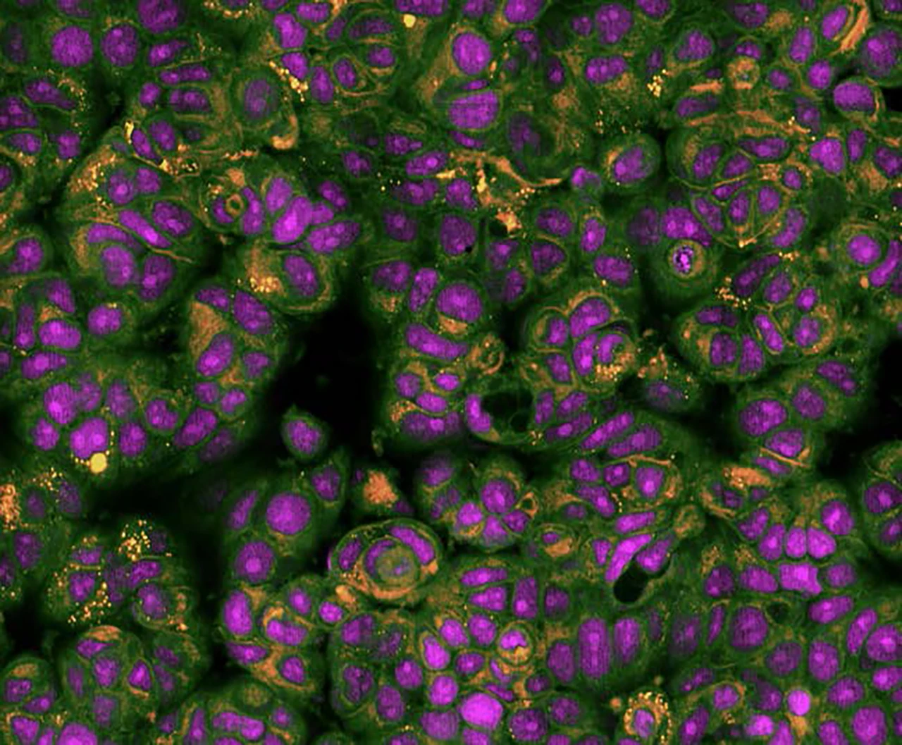
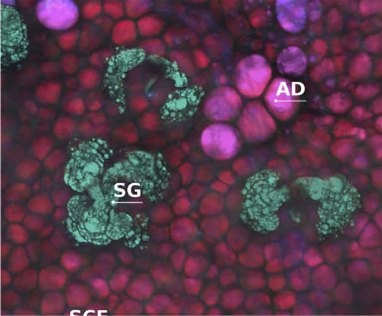
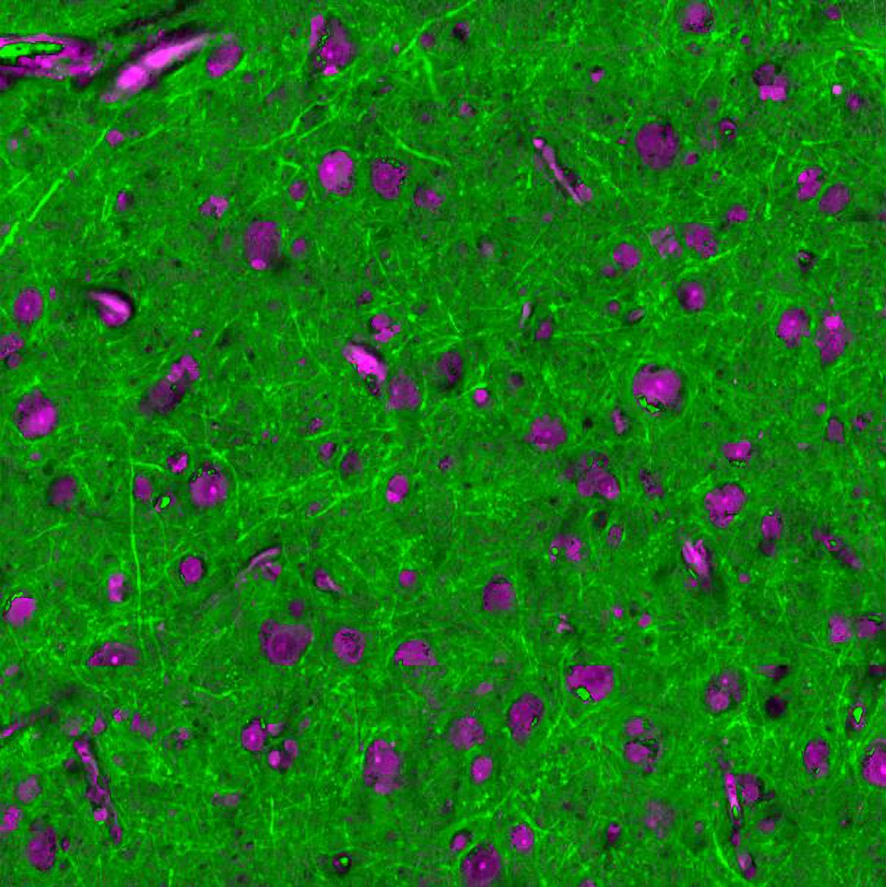
SRS reveals cellular and tissue processes that remain invisible to fluorescence microscopy. It highlights lipid droplets, membranes, and cytoplasmic regions based on their natural molecular composition. This chemical contrast allows to study metabolism and membrane organization under physiological conditions.
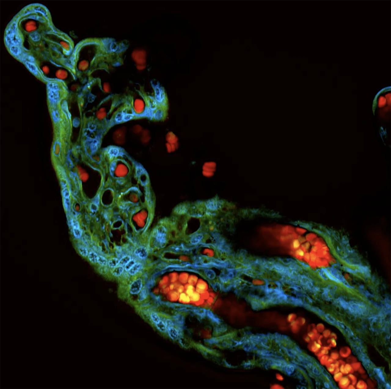
The multiphoton nature of SRS enables imaging deep inside thick and unprocessed tissue. It reveals how lipids, proteins, and blood components are organized within intact organs such as the placenta, preserving their natural architecture and biochemical context.
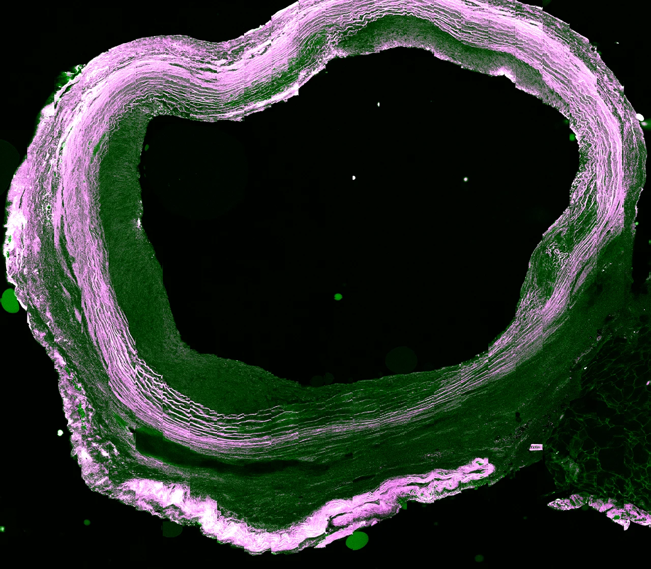
SRS combines structural and molecular information in a single, integrated view. It distinguishes collagen fibers, lipid domains, and small calcifications within tissue, revealing the chemical architecture that defines its morphology.
SRS visualizes molecular activity in living tissue without fluorescent labels. It captures how unlabeled drugs penetrate through skin and interact with lipid-rich layers, allowing molecular transport to be studied in real time under natural conditions.
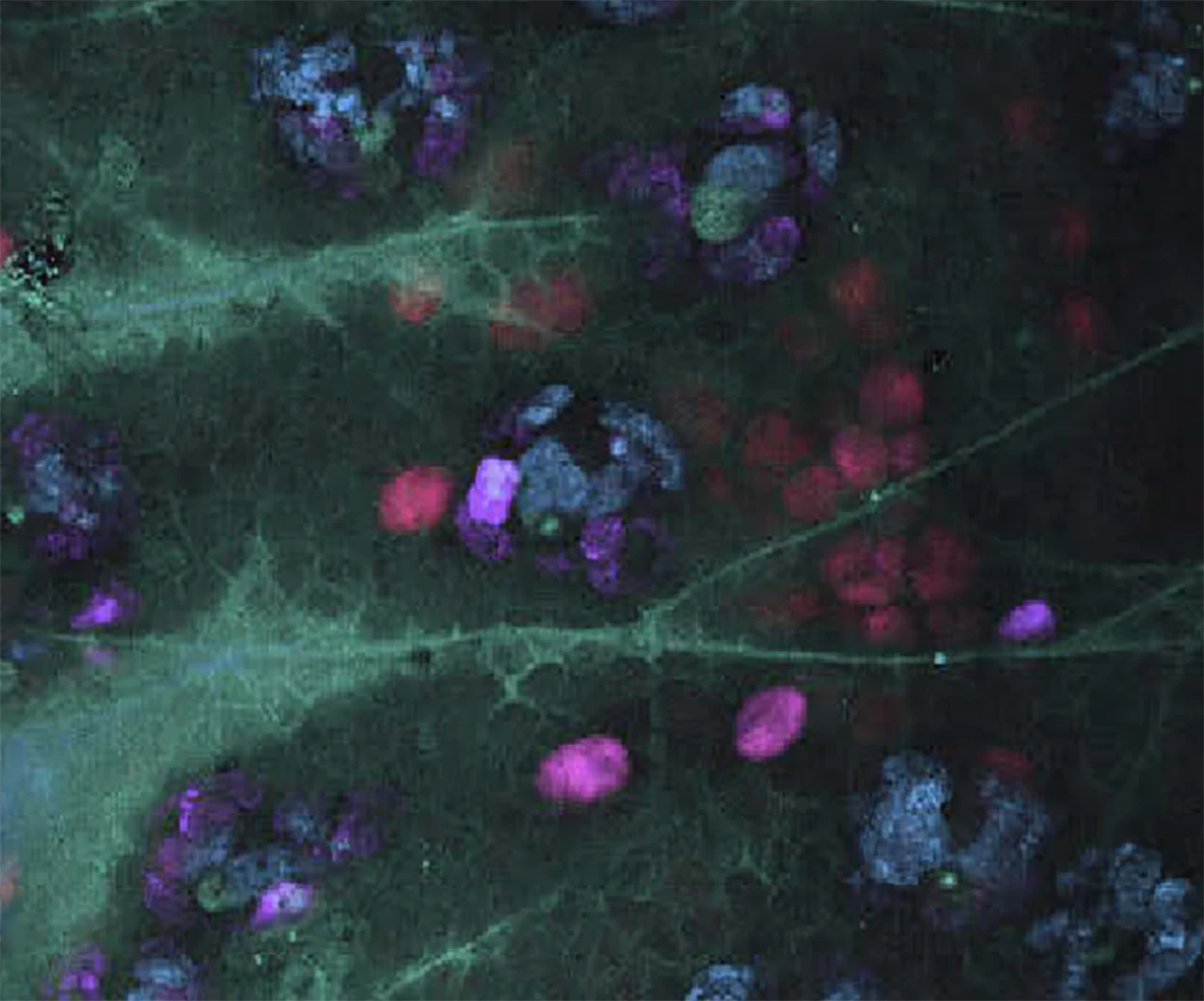
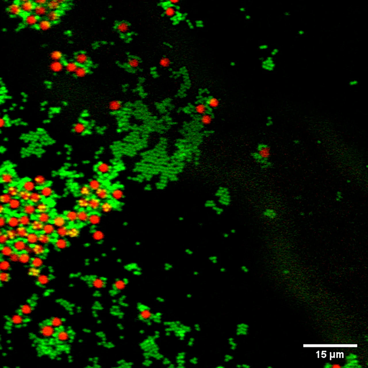
SRS reveals biochemical patterns that remain invisible to conventional microscopy. In brain tissue, it highlights lipofuscin accumulations and other molecular features, exposing the chemical composition behind structural changes.

SRS distinguishes materials based on their unique vibrational spectra. Different types of microplastics or chemical compounds can be identified instantly, demonstrating the versatility of molecular imaging beyond biological research.
Instant insight
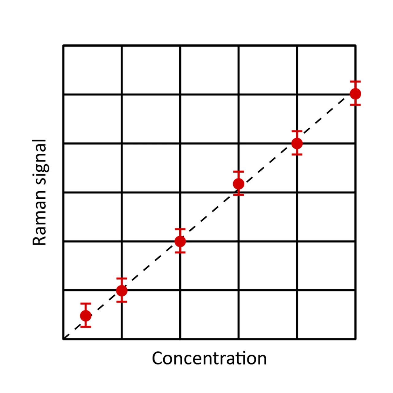
SRS provides the benefits of Raman spectroscopy without its limitations. It generates no fluorescent background, works with standard glass slides, and produces a linear signal that directly reflects molecular concentration. This enables quantitative analysis of chemical changes such as metabolism or drug uptake.
Molecular context
With a complete molecular spectrum in every pixel, SRS turns complex cellular reactions into clear, analyzable information. Changes in molecular composition and spatial organization reflect cellular dynamics and genetic alterations, linking structure and function and enabling analysis of cell states and compound effects.
Use cases
Our versatile laser technologies deliver precise, label-free imaging across many fields, improving healthcare globally.
Lets you watch how cells act and interact in real time.
Helps analyze fat processing inside live cells without labels.
Tracks how drugs move and change within cells over time.
Reveal molecular and structural changes relevant to cancer biology.
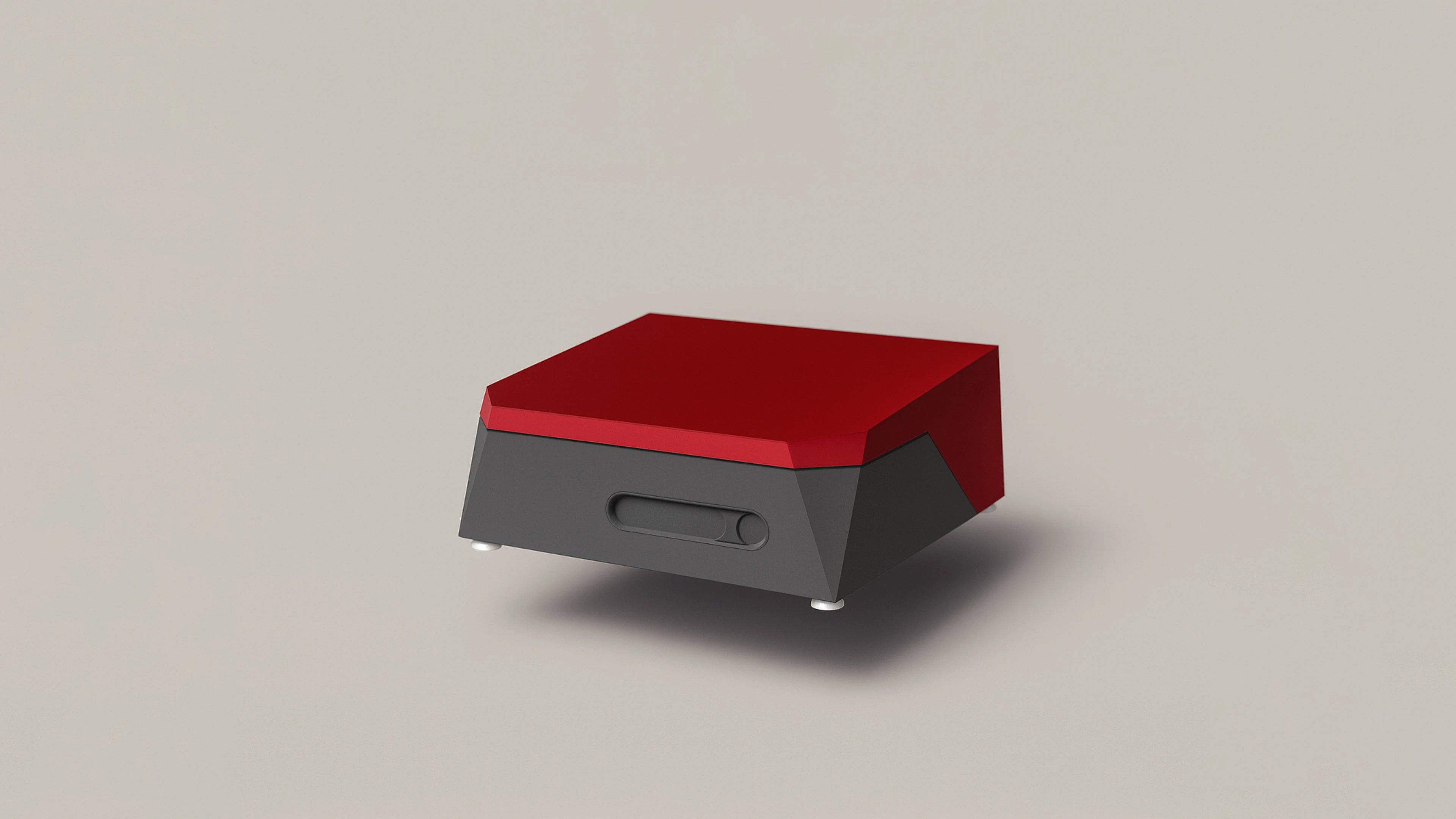
Products
Step into technology that surpasses conventional systems and unlocks the future of discovery and diagnostics.
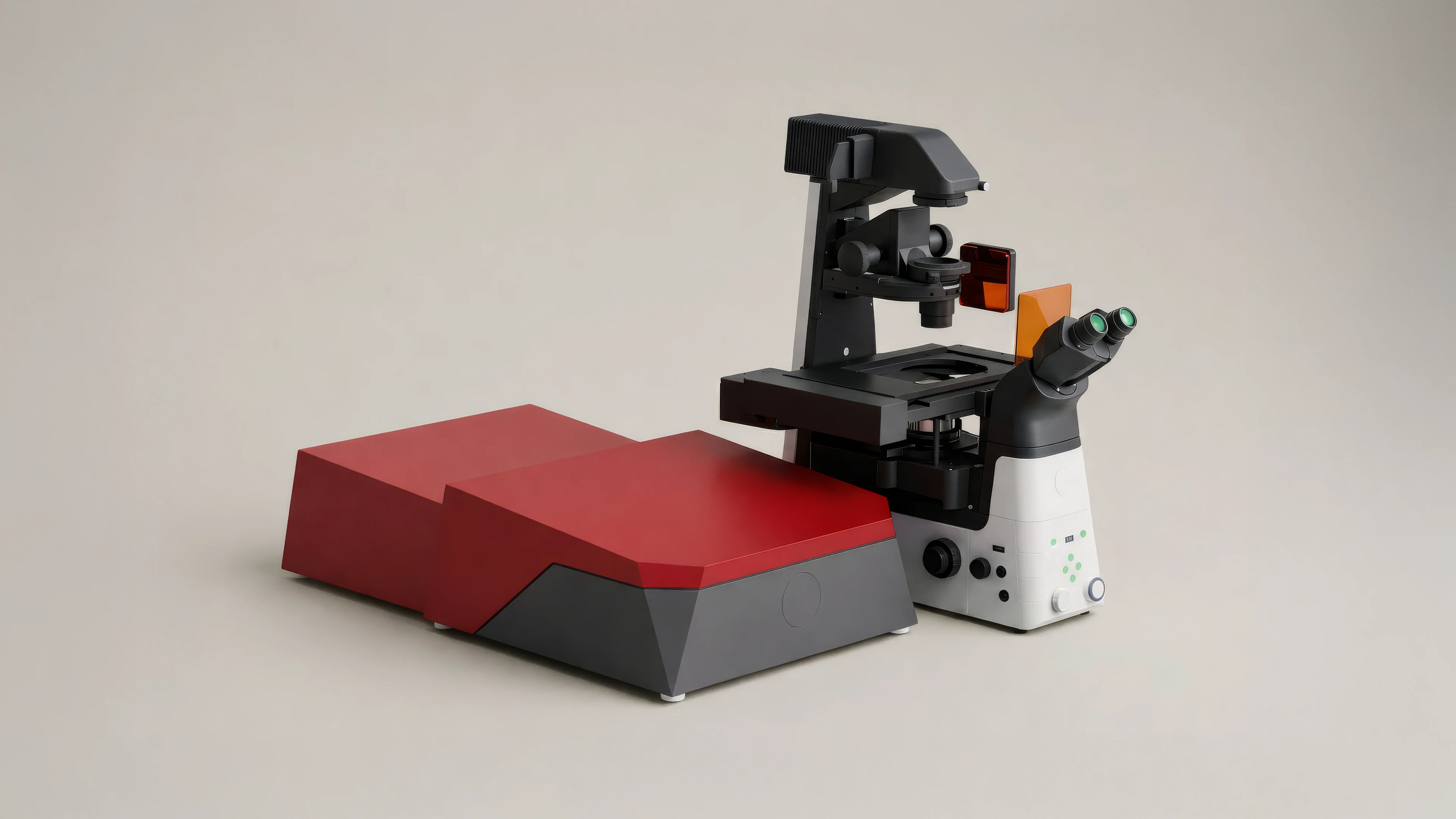
Our Noctua is an automated, label-free SRS system for fast chemical imaging with easy setup, full spectral access, and rapid tuning – ideal for pathology and live cell studies.
Portfolio
Our products provide powerful SRS imaging solutions, from flexible lasers for microscopes to a fully automated, label-free system with minimal setup.
Fully automated microscope system
SRS & CARS imaging
TPEF & SHG ready
Easy plug & play operation
Computer-controlled tuning
Air-cooled
Balanced detection
Live spectrometer
Automated temporal beam overlap
Pre-adjusted spatial beam overlap
Individual beam divergence control
Polarization control
Automated power control
Trigger in and out for precise synchronization
Integrated laser scanner
Fully automated
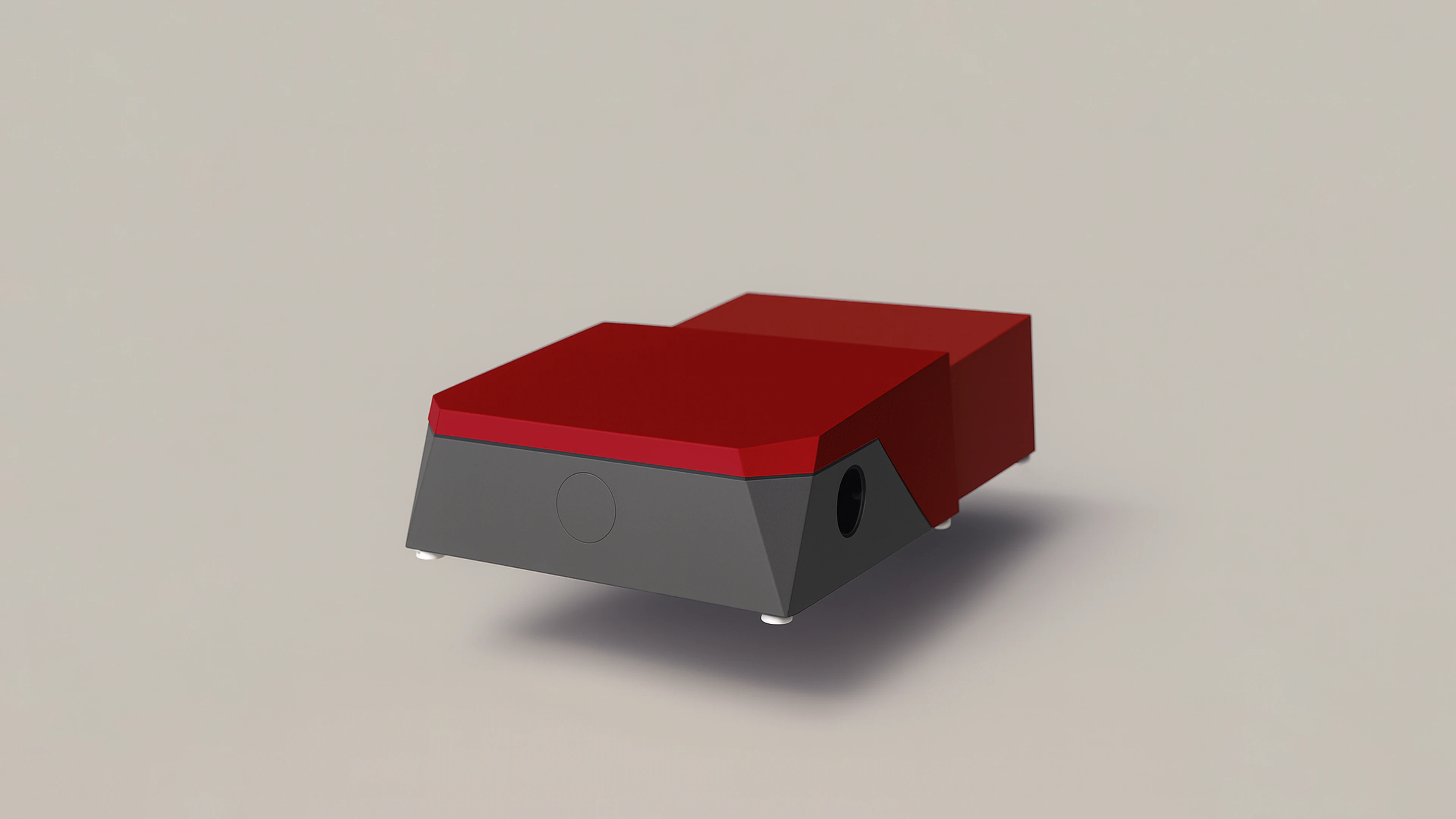
Compact laser platform for SRS imaging
SRS & CARS imaging
TPEF & SHG ready
Easy plug & play operation
Computer-controlled tuning
Air-cooled
Balanced detection
Live spectrometer
Automated temporal beam overlap
Pre-adjusted spatial beam overlap
Individual beam divergence control
Polarization control
Automated power control
Trigger in and out for precise synchronization
Integrated laser scanner
Fully automated
Compact dual-output laser system
SRS & CARS imaging
TPEF & SHG ready
Easy plug & play operation
Computer-controlled tuning
Air-cooled
Balanced detection
Live spectrometer
Automated temporal beam overlap
Pre-adjusted spatial beam overlap
Individual beam divergence control
Polarization control
Automated power control
Trigger in and out for precise synchronization
Integrated laser scanner
Fully automated
Fast wavelength switching across a wide tuning range is a defining feature of Refined Lasers and supports Raman imaging experiments beyond the reach of conventional laser sources.
Prof. Conor Evans
Associate Professor, Harvard Medical School, Wellman Center for Photomedicine, Massachusetts General Hospital
Contact
FAQ
Quick answers to common questions.
Content.
Longer content.